richard.savage@LIGO.ORG - posted 11:14, Tuesday 02 June 2015 - last comment - 11:42, Tuesday 02 June 2015(18773)
Pcal beam localization camera images at Xend
NutsineeK, RickS
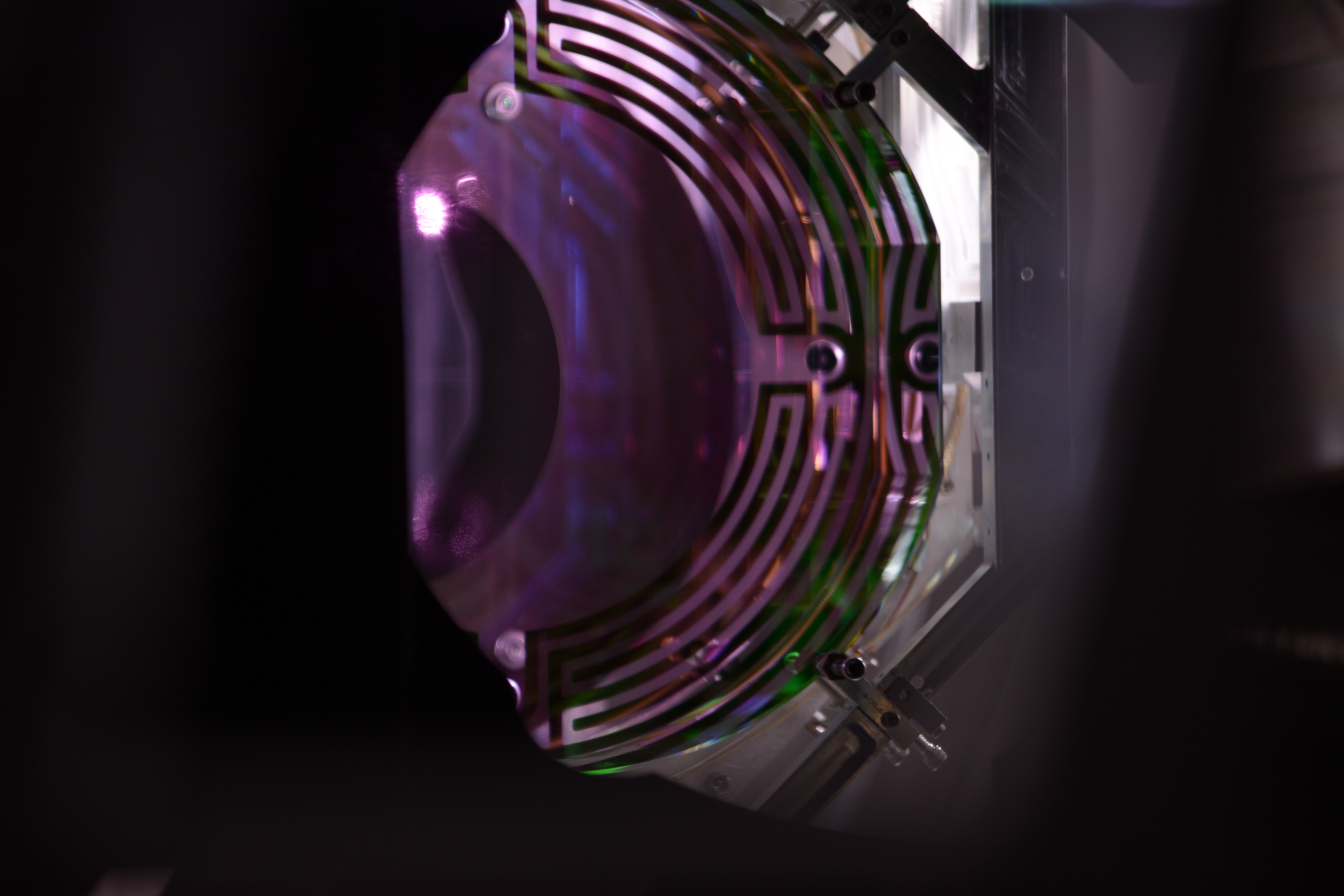
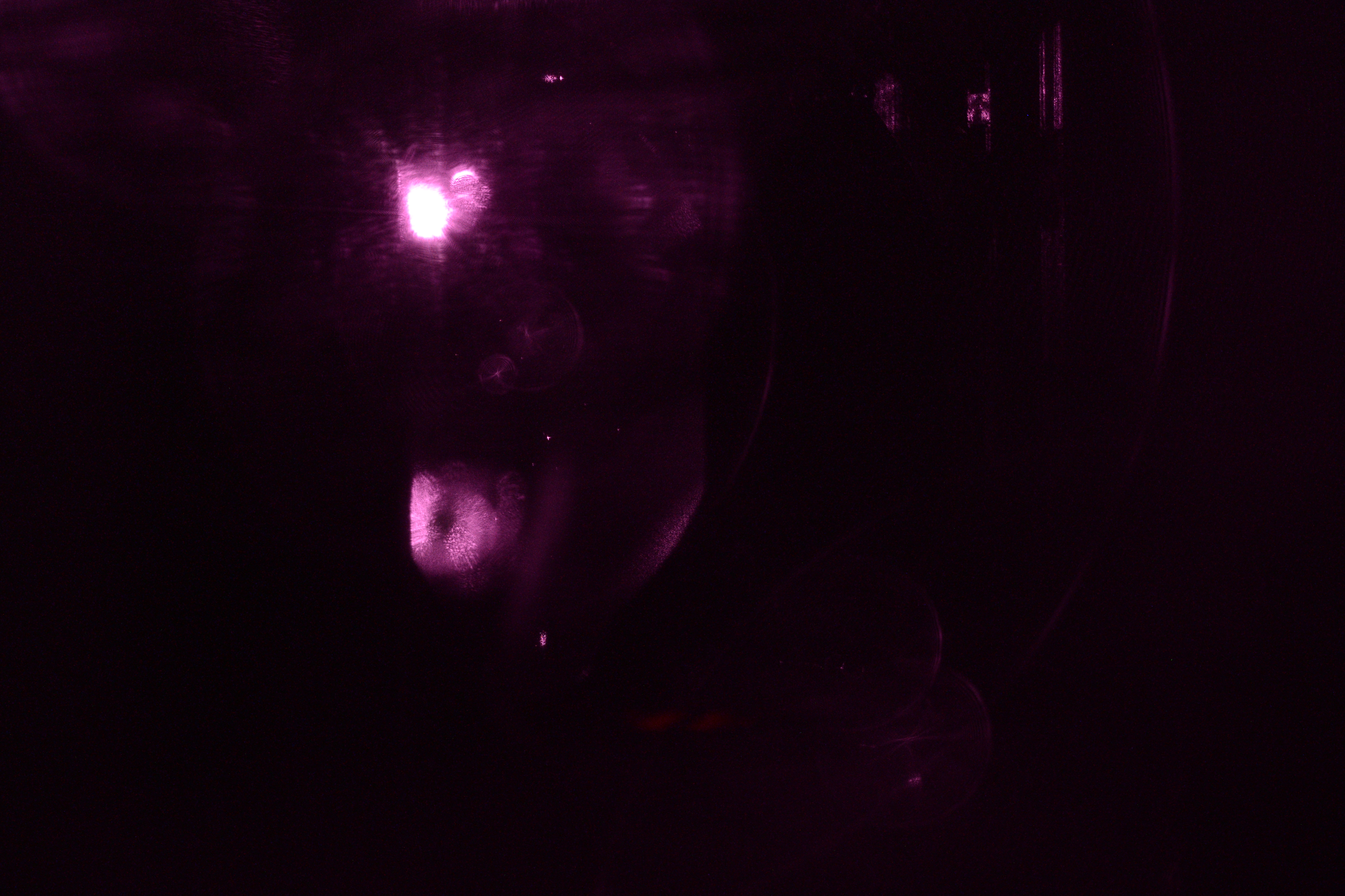
This morning, we went to Xend to capture images of the ETM with the illuminator on and the green ALS and red OptLev beams blocked.
The procedure for capturing the measurements is as follows:
- Turn off Pcal excitations on the PCal medm screen.
- Note the OFS Offset level (6.0 for LHO Xend)
- Set OFS offset to maximum level (10 at EndX)
- Close ALS green ALS light shutter
- Block the optical lever beam with the Pcal red aluminum beam block. Need to remove the 8-32 screw from the strip that blocks the slot in the viewport protector and remove the strip (be careful not to drop the screw).
- Remove the Pcal Rx module cover and block the Pcal beams at the entrance to the Rx PD integrating sphere using a razor blade dump.
- Take two images of the ETM, the first with the illuminator on and focused on the edge of the ETM (visible) and the second without the illuminator and focused on the Pcal spots on the ETM surface (infrared).
- Set the OFS offset back to the nominal level.
- Turn on the excitations.
- Remove the beam block from the Optical Lever and replace the strip that covers the slot in the viewport protector.
- Open the shutter for the ALS green beam.
- Remove the beam dump from the Pcal Rx PD in the Rx module and replace the cover.
The images will be attached to this report shortly.
Comments related to this report
Images attached to this comment